|
Observations and speculations from an undergraduate class of NON-insect experts
(because as scientists, why shouldn't we try to figure things for ourselves!):
SPIDER
 |
|
 |
| Appearances to the contrary, the spider is really only about 1 mm end to end (see scale bar at the bottom left). |
|
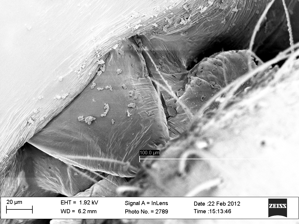
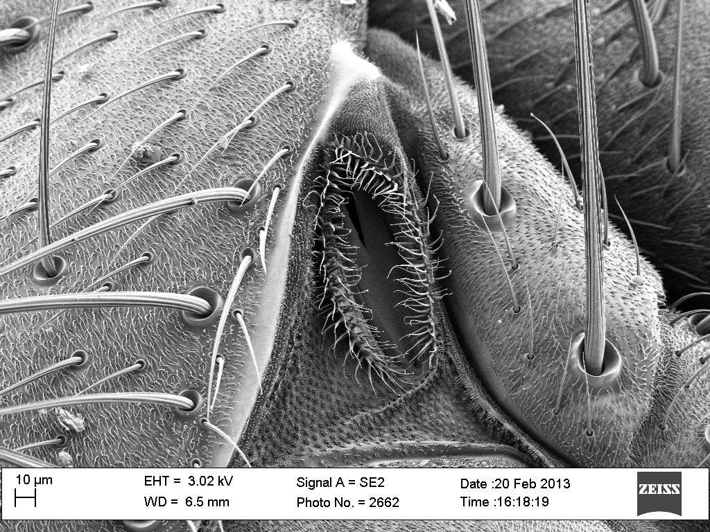
Spiders have hard exoskeletons mimicking suits of armor. At their flexible joints, suits of armor employ small protective flaps. Is this a similar flap protecting the spider's waist?
|
 |
|
 |
| But the elbows (leg joints) do not seem to have protective plates. Instead the gaps are filled with small regions of flexible tissue (just as you see at a crab's leg joints). |
|
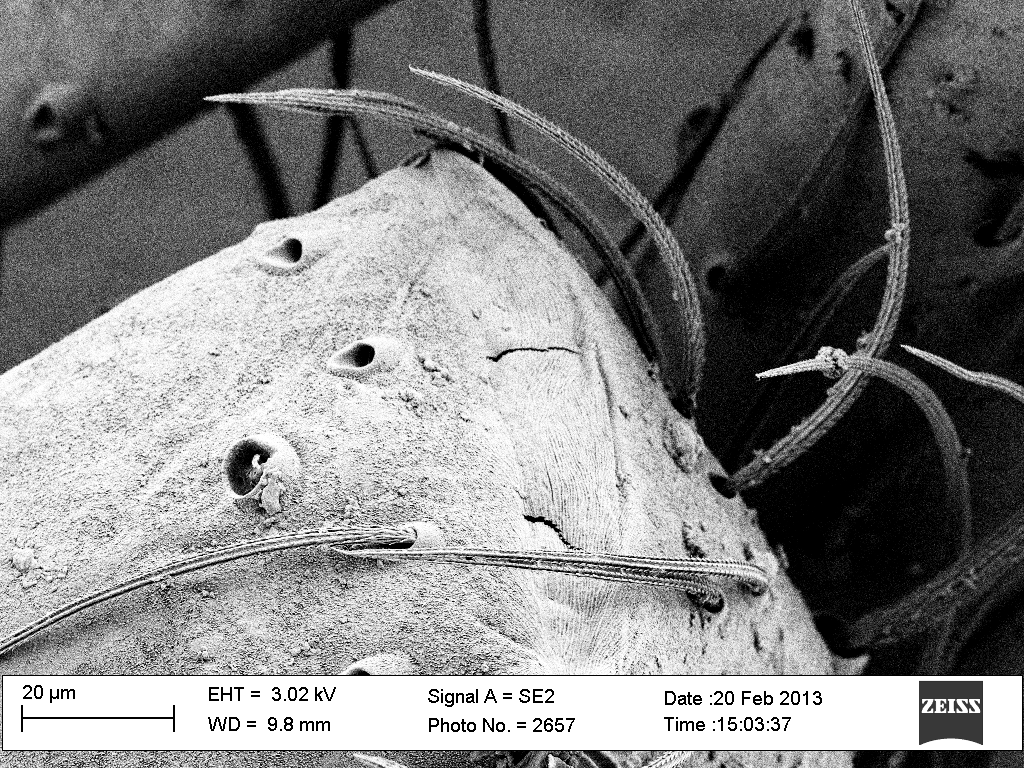
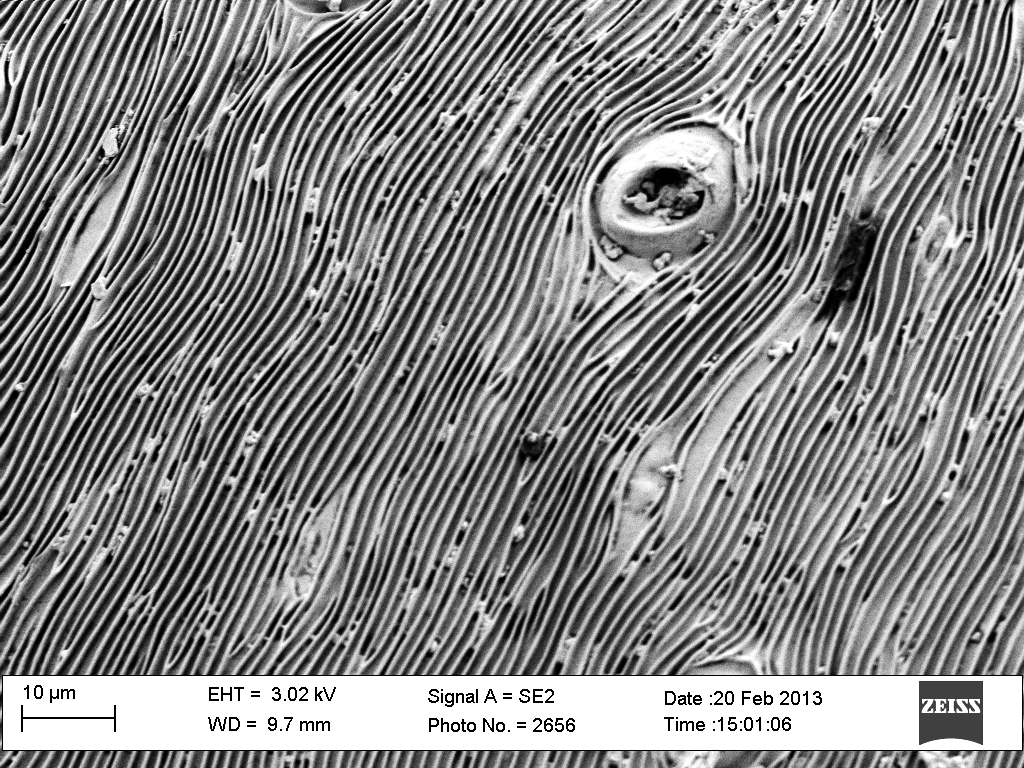
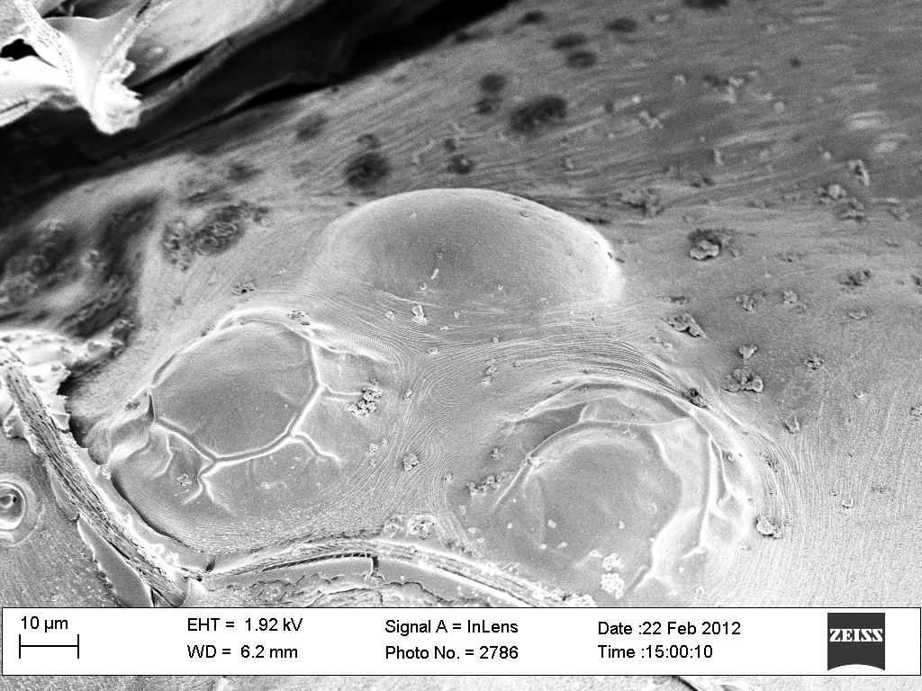
Are these breathing holes all over the back of the spider's abdomen?
And while the surface of the head was moderately smooth, the abdomen was covered by these corrugations. Might they allow the abdomen to expand and contract as the spider eats and excretes?
|
 |
|
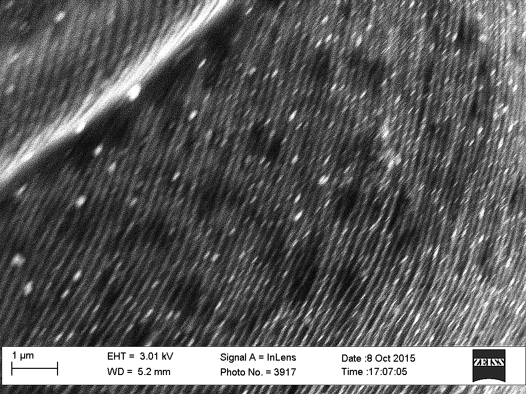 |
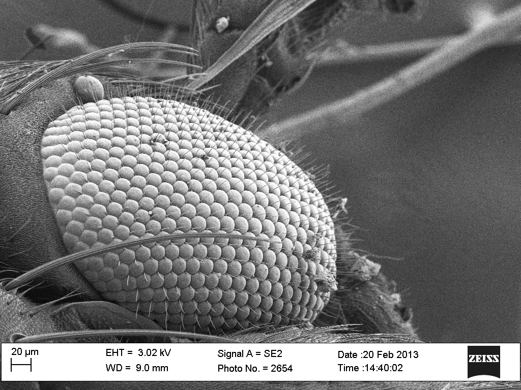
Here are three eyes on one side of the spider's head (after drying out for almost a year). At very high magnification the surfaces appear to be covered with ridges (shown enlarged in the micrograph to right). We wondered why these ridges did not distort the spider's vision. But then we noted their scale: they were far narrower than the wavelength of visible light (0.4 - 0.6 microns), so light would not be scattered by this roughness.
|
|
Then we noticed that the ridges on each of the set of three eyes ran in markedly different directions. Could these nanoscopic ridges act as light polarizing filters? If yes, that would allow each eye to see with differently polarized light, perhaps better revealing the spider's environment or prey. |
 |
|
 |
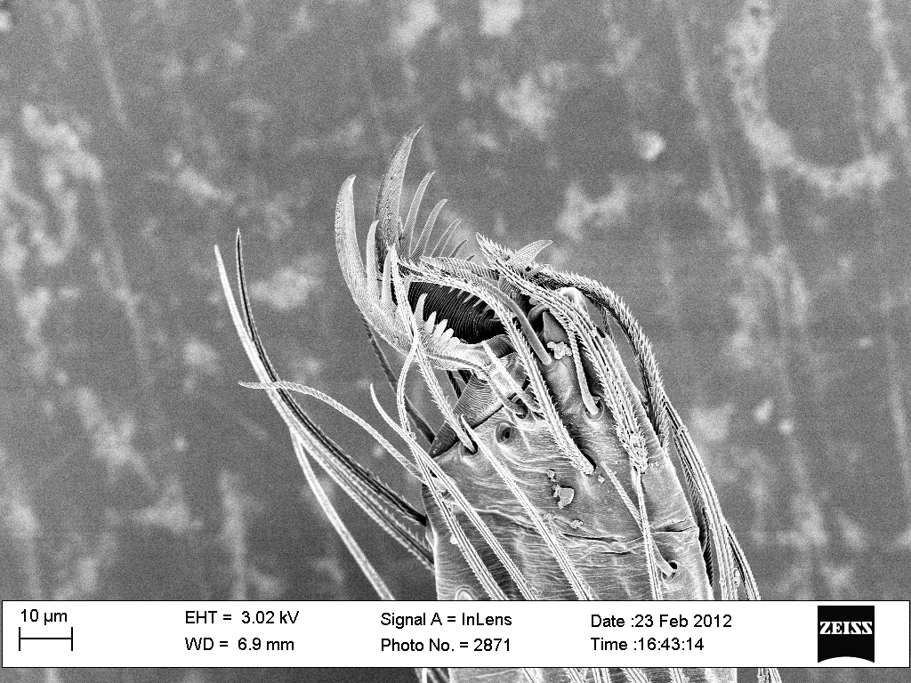
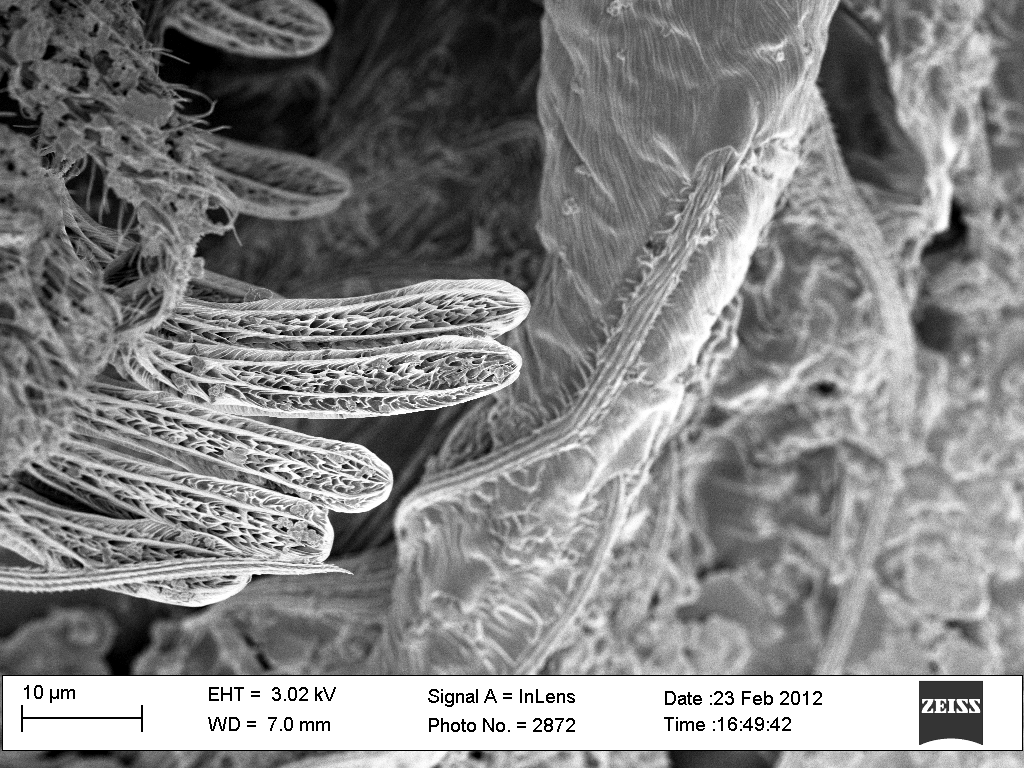
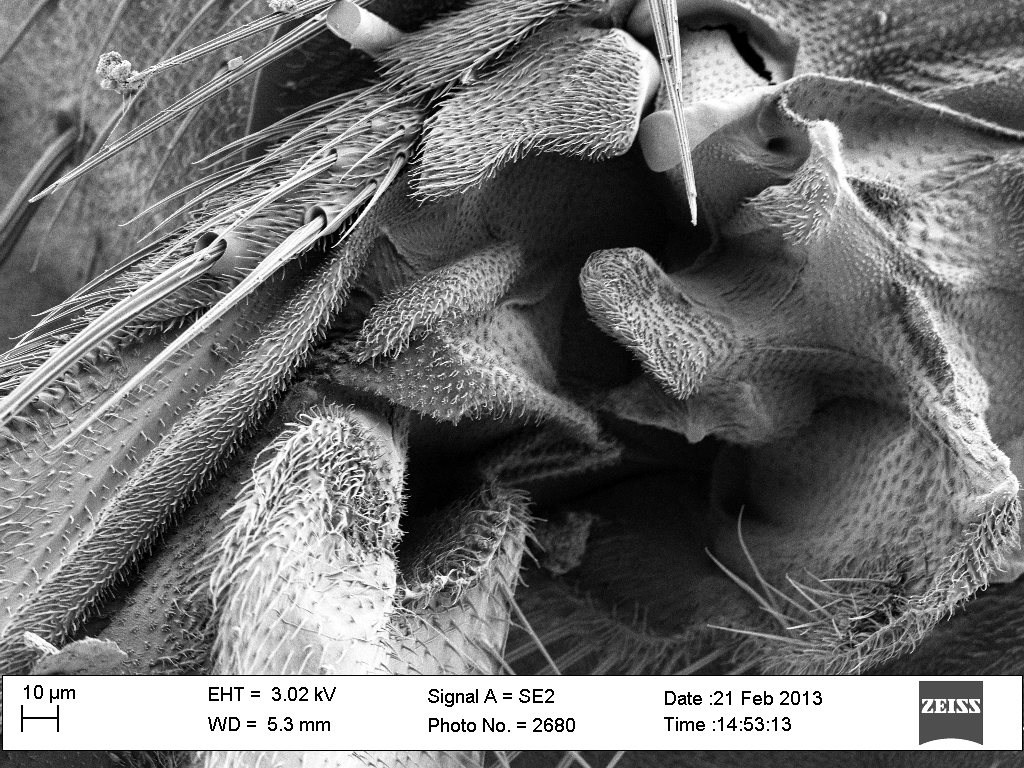
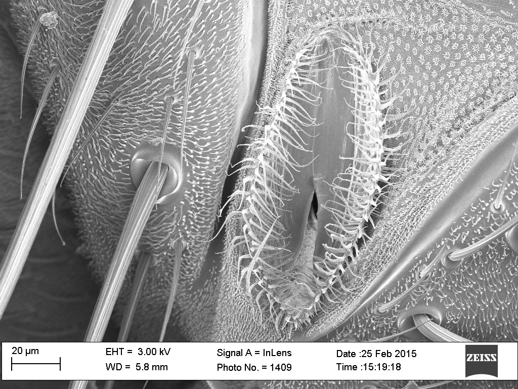
| Here is one of the spider's toes showing two opposing almost hand-like combs in which the spider may grasp the filaments of its web. |
|
Away from the toe (to the right) the spider's skin/shell has its typical, more or less smooth, texture.
But everything AT the toe has a very pronounced ribbed texture. Is this a way of minimizing the contact area with the silk filaments, making it less likely that the spider will get stuck to its own web?
|
 |
|
 |
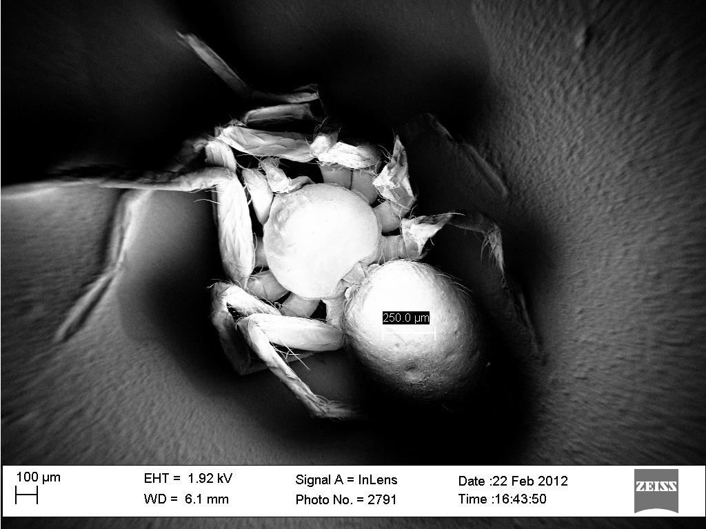
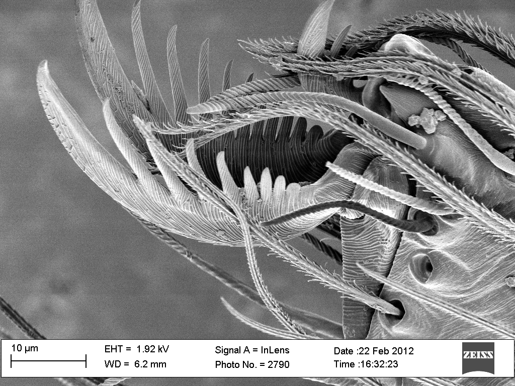
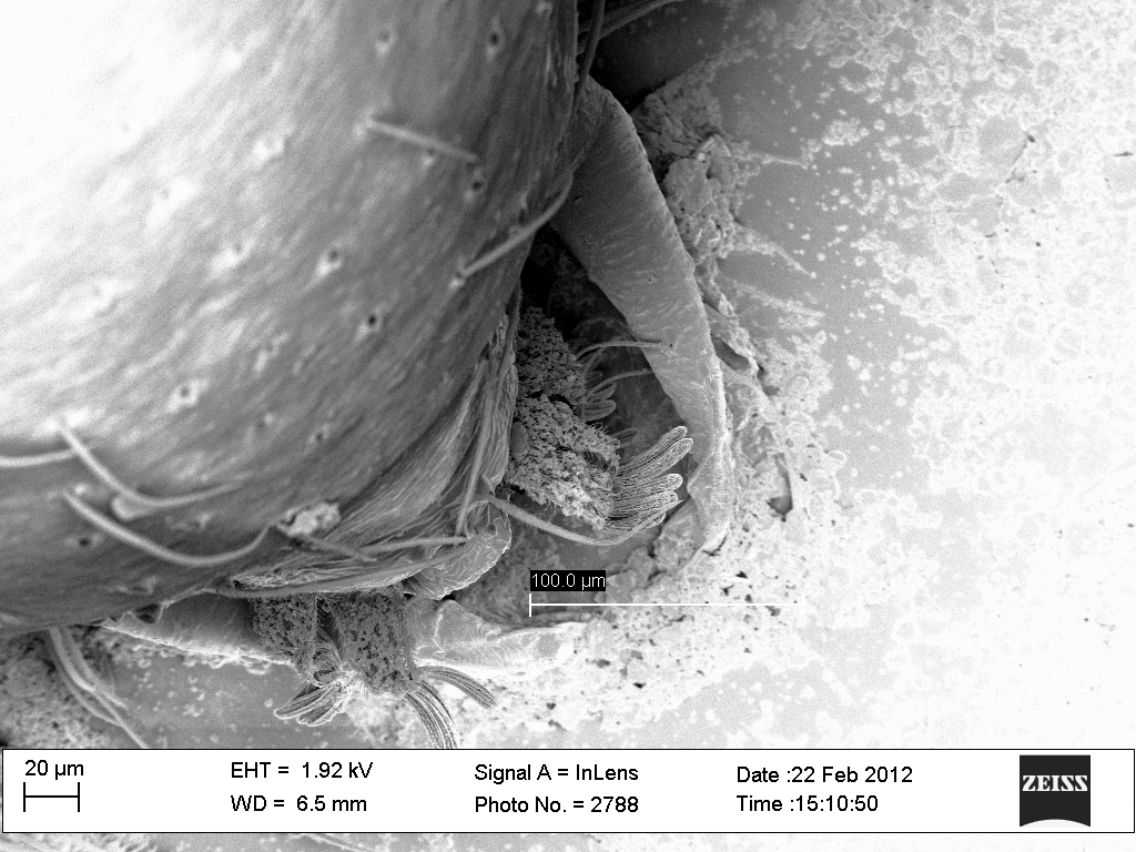
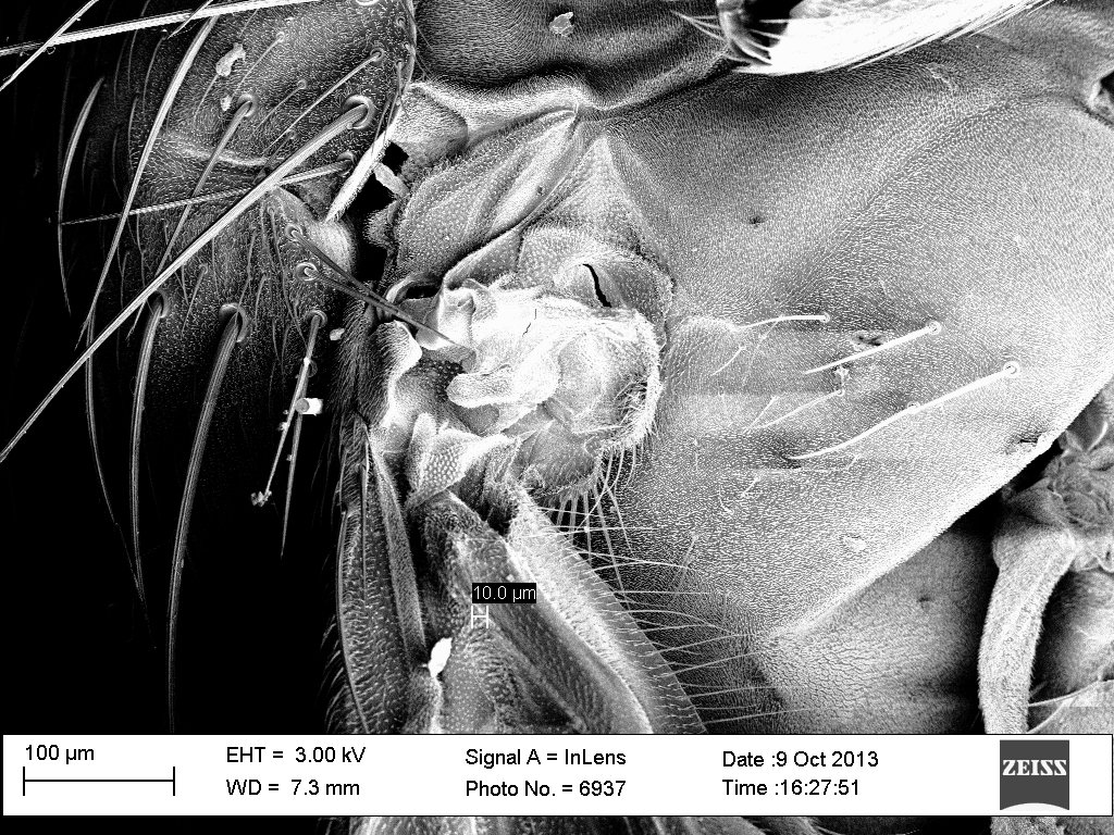
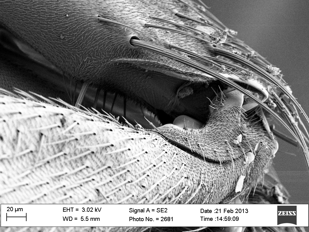
Here it the spider's rear end showing what we assume are the spinnerets plus the small arms used to pull out the silk.
|
|
The liquid silk "dispensers" themselves - employing surface tension to suck the liquid silk up into their partially hollow cores? |
MOSQUITO
 |
|
 |
The mosquito's eye. Are the fine hairs between each and every lens protection against parasites? Or, by holding droplets away from the surface, do they help the mosquito to shed water?
|
|
The end of the mosquito's blood sucking proboscis? The size looks right in that the apparent opening at the top is big enough (~ 15 microns) to allow ~ 4 micron diameter human red blood cells to pass through. (It had broken off the head so we could not be totally sure).
|
 |
|
 |
The incredibly complex shoulder joint of the mosquito (the wing extends out towards the lower left).
|
|
The shoulder joint seen more closely (viewpoint rotated 90 degrees CW from one at left).
|
 |
|
 |
Mosquito nose? Here you can see the fine texture of a antenna structure above eye. Note the thousands of nanometer scale holes.
|
|
This was just forward of the mosquito's shoulder/wing joint. An ear? A breathing portal? Something else? |
 |
|
 |
Another view of the thing just forward of the shoulder/wing joint
|
|
The surface of the mosquito's wing, which like all other surfaces was covered by fine hairs. Again, are they parasite or water repellers? Or might they play a role in setting up an aerodynamic boundary layer helping the wings to beat faster?
|
 |
|
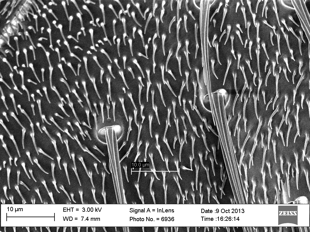 |
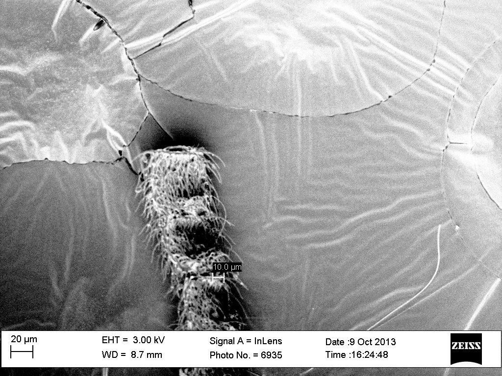
| Mosquito knee. |
|
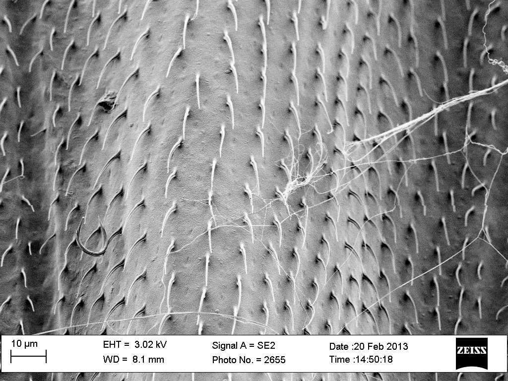
Hairs on the mosquito's abdomen |
MORPHO BUTTERFLY WING
 |
|
 |
We had read about many nanoscience researchers taking interest in the wings of the morpho butterfly. But the articles were frustratingly vague about WHY they were interested! We suspected it might have something to do with the strange shimmering blue color of these wings. The fact that color changed with angle suggested that the wings might be covered with diffraction gratings (i.e., parallel ridges separated by fractions of a light wavelength). |
|
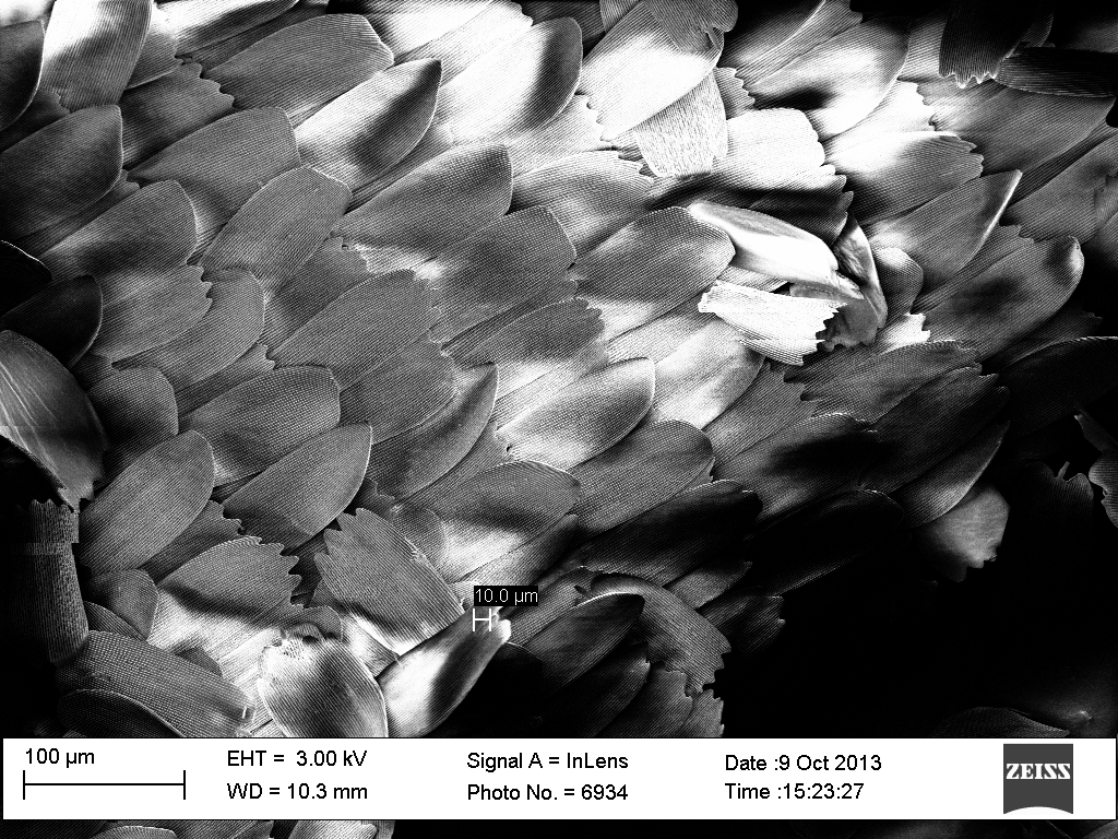
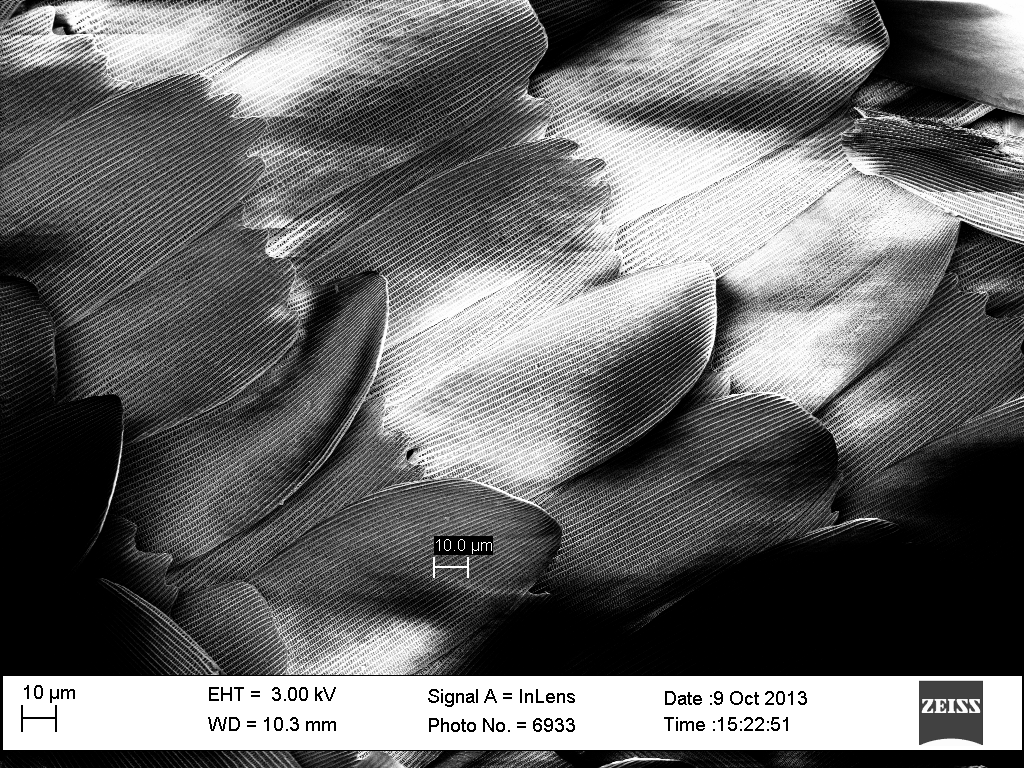
Then Luke came up with a specimen. At low magnification the wing appeared to be covered by these petal like structures.
|
 |
|
 |
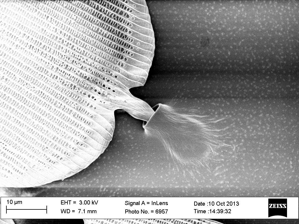
Here is a region where the petals were farther apart. At the bottom center, you can see how the petals are attached to the wing by a tiny stem at their left end. At the right edge of the picture one of the petal stems has pulled free, leaving its empty socket to the left.
|
|
Here is an enlargement of a stem still in its socket.
|
 |
|
 |
Here it is again at even higher magnification.
|
|
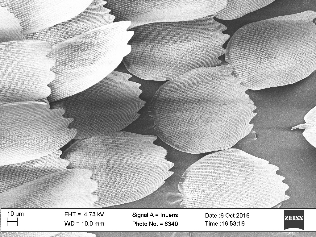
And here, one of the petals has pulled loose, flipping over to reveal that its backside is smooth.
|
 |
|
 |
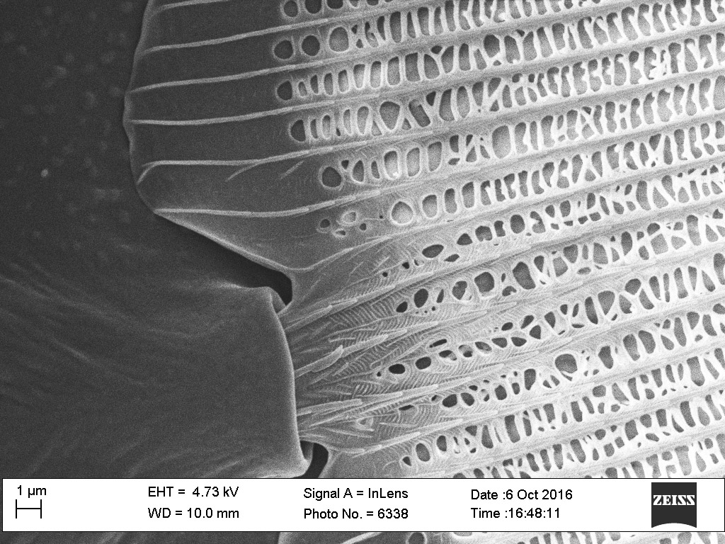
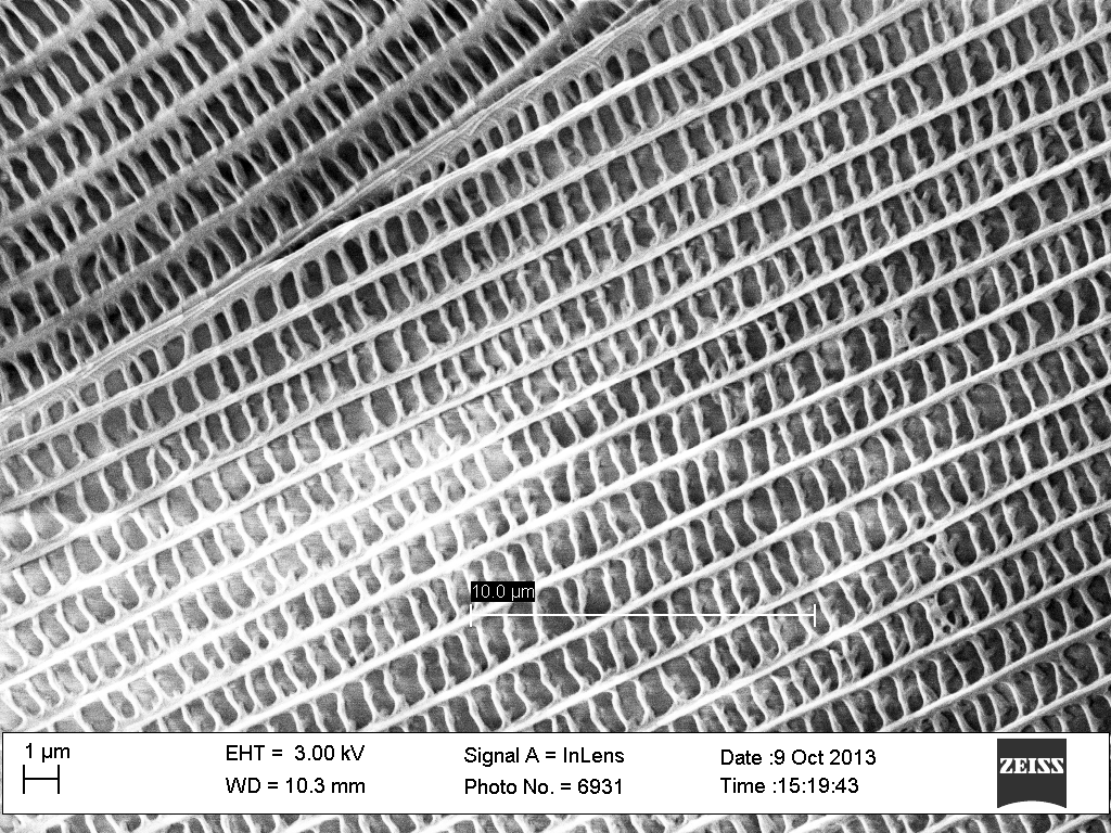
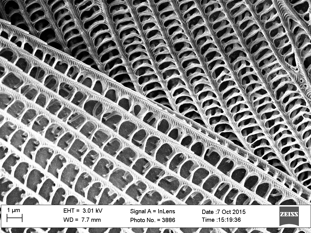
| But the frontsides are anything but smooth: They are covered by rectangular grids. |
|
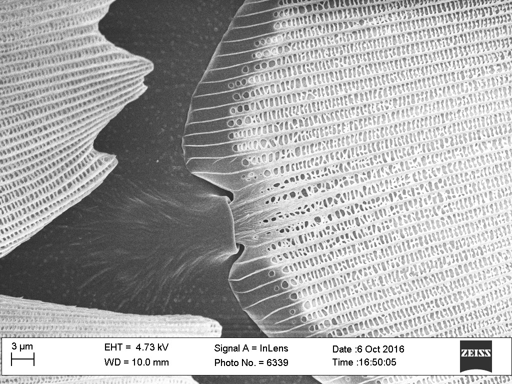
Which, at even higher magnification, are revealed to be trellis-like structures suspended ABOVE the solid surface of each petal.
|
 |
|
 |
On different petals, the rectangles had different widths. Petal to petal, the widths varied from about 1.5 microns down to 350 nanometers. This would correspond to light wavelengths from the infrared to ultraviolet. But on most petals, cell widths had sizes corresponding to visible light wavelengths (i.e., 400-600 nanometers), and the most common widths were about 400 nm (the size of blue light). This would tally with the fact that we perceive these wings as having a shimmering royal blue color.
|
|
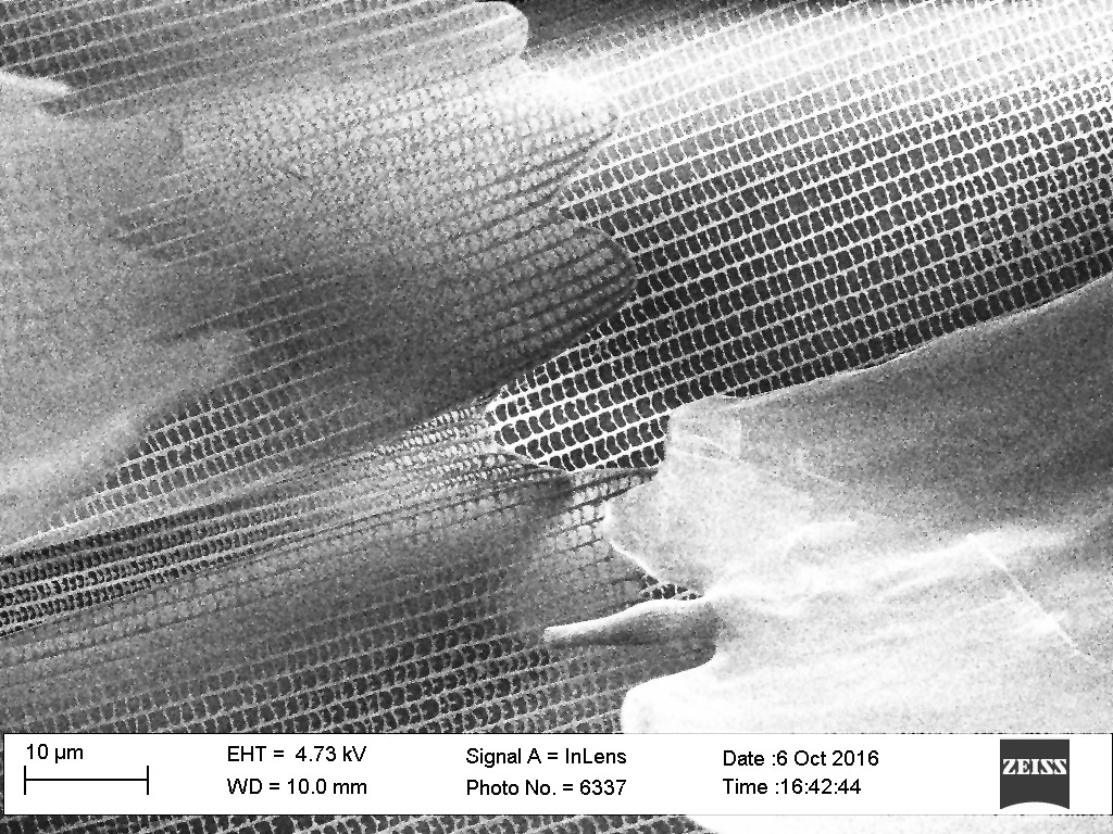
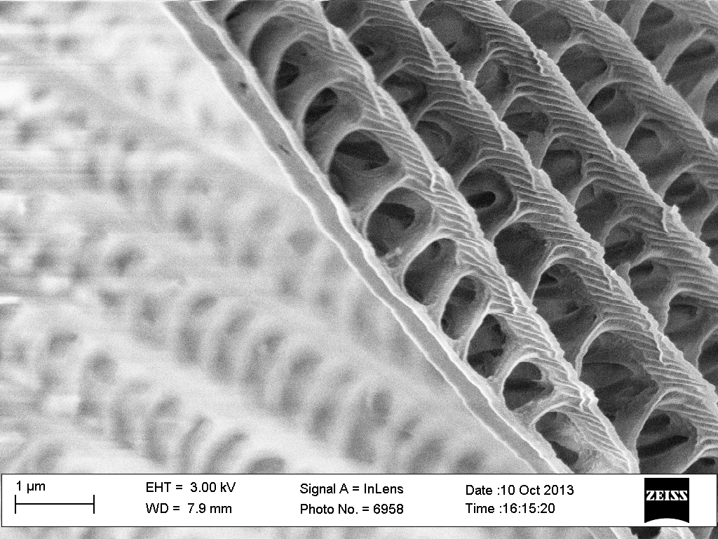
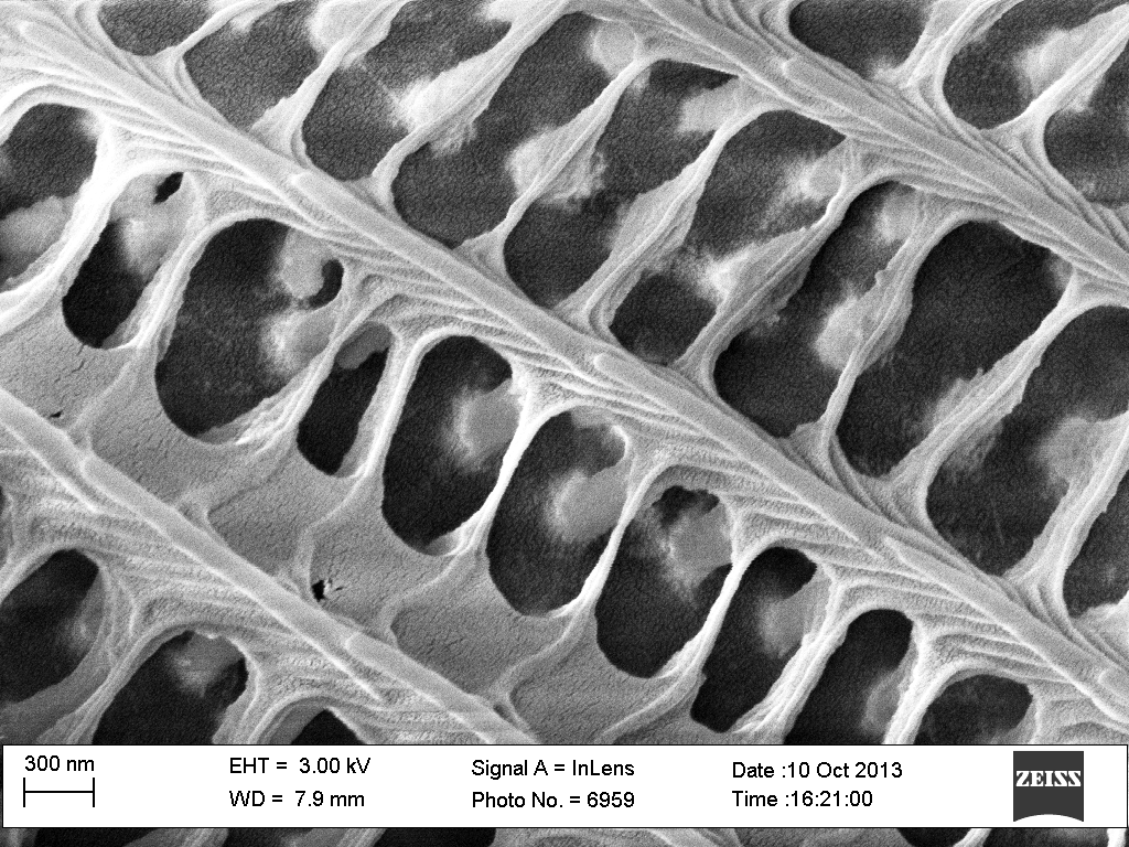
Here you can see the incredible nanostructure of these "petals." On their surfaces they appear to have lattices suspended atop pillars, with the finest features or the lattices well under 100 nm in width.
The sizes and regularity of these rectangular lattices supports the idea that with these "petals" the morpho butterfly is indeed using diffraction gratings to direct specific reflected colors into specific directions. |
 |
|
 |
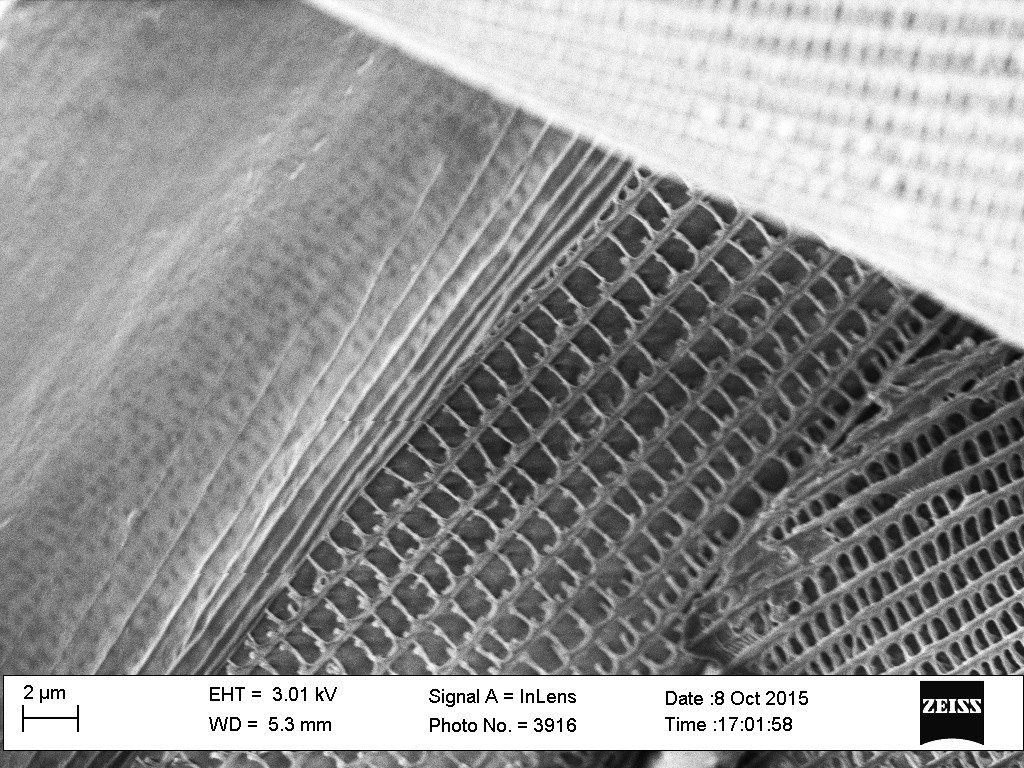
Here is a closeup of a region of the wing that was roughly handled during sample preparation. The petals at top and bottom right are intact. But left and center seem to be a petal that has been torn apart: The more or less smooth underlying petal base is visible at left. The lattice torn from it, now upside down, is beside it at the picture's bottom center.
|
|
But what evolutionary purpose could this use of diffracted light serve?
Zooming back out, you can again see how the "stem" of each "leaf" is attached to a "socket" in the wing's surface. Could this arrangement allow the butterfly to deliberately twist the "petals?" If so, the diffracted light beams would then be swept out in different directions.
This might produce a strange shimmering light pattern that could mimic the way squids use chromophore-based colors to flash confusing patterns at pursuing predators (or, of course, it could just be a peacock like way of attracting mates).
|
Copyright: John C. Bean |