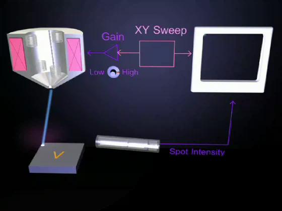
Here the amplifier is set to high gain. This setting scans the electron beam over a large area.
Exaggerating a bit, imagine that the blue stage is made of a material that emits very few electrons, while the "V" emits many. Then every time the beam passes over the V there is a flash of electrons. These are collected and amplified by the detector causing the spot on the CRT screen to brighten. Then, because the CRT spot follows a synchronized path, we get an image of the V on the screen.
The image is thus produced by the pattern of the target's electron emission.